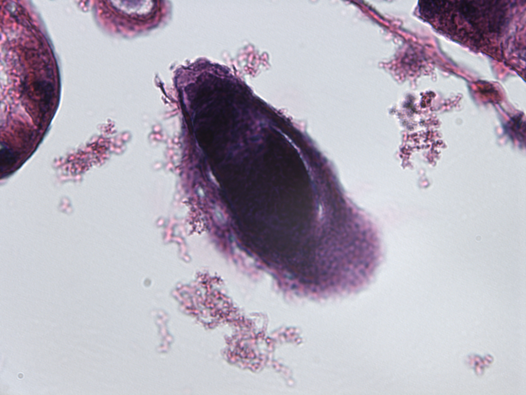
Histological transversal section (17 µm, counterstained by eosin and hematoxylin) of a Sesamia nonagrioides larva (L4) parasitized by Cotesia typhae. In the center, a parasitoid egg (dark purple) is surrounded by host immune cells, forming a defensive capsule which will kill the egg and end the parasitism process.